1. The name of the pumping organs of an earthworm are the five aortic arches, which is more commonly referred to as hearts.
A heart is located in the dark mass of blood and organs.
2. The worm's digestive tract goes as follows :
Mouth: The opening to the digestive tract
Pharynx : Helps suck the food in
Esophagus : Moves food to the crop
Crop : Moistens food
Gizzard : Grinds up the food ( Mechanical digestion )
Large Inestine : Further digestion and absorption ( Chemical digestion )
Anus : Excretes waste
 Here is a picture of a worm fully cut open. You can see the different organs of the worm and the path that it takes.
3. The part of the earthworm that serves as its brain is the cerebral ganglion. It is connected to the rest of the body through a ventral nerve cord.At each segment of the ventral cord is a ganglion which is a swollen region of nerves that connects every segment to the brain.
 Here is a close up image of the worm's brain.
4. The parts that are included in the worm's excretory system are the anus and nephridia.
 A picture of the lower half of the worm. ( Where the excretory system is located )
5. You can find out what a worm eats if you take a look at its digestive system. If you dissect the worm, you can actually find soil that was traveling through its body.
 Dissecting the worm can show you its diet.
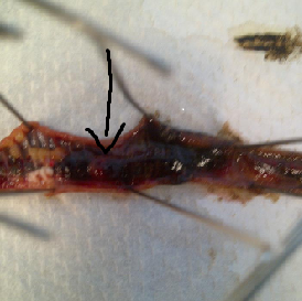
6. The setae found on an earthworm are tiny bristle like structures that assist in movement. They help by latching onto terrain so predators cannot pull them away..
 I was unable to take a picture of the setae so instead, I found a picture that the setae is clearly visble.
7. The earthworm's crops store the worm's food as it comes down the esophagus and the gizzard grinds it down. This process makes the worm well suited to its environment.
 Here is a picture that shows the esophagus and gizzard.
8. If I cut past segment 32, I would see the rest of the digestive system, nephridia and the worm's food.
 This picture shows a portion of the bottom half of the worm. It shows the digestive system and some nephridia.
9. When earthworms start to reproduce, they both exchange sperm with one another because they are hermaphroditic. Both worms would then proceed to collect the sperm in special sacs where it will stay there until it is mature. Once this happens, the worm's clitellium begins to create a sticky substance that will form a ring which contains the now fertilized eggs.
 This picture shows the worm's seminal vesicles.
Purpose-4/4
ReplyDeleteConnection to class-2/4
Personal Reflection-4/4
Conventions-4/4
Requirements-9/9
23/25