Friday, January 25, 2013

Rat Dissecrtion:

          Last Thursday, our Biology class was fortunate enough to participate in a rat dissection. Upon arriving to class, we were divided up into groups of three or four where we discussed roles in our dissection. After a few minutes, all of the groups received their rats and we began examining its external features. Our group was given a female rat, but unfortunately, our rat was not pregnant at the time of death, so we were unable to see any rat  fetuses. Our group was able to analyze its whatever we could see to find out some information about the appearance of the rat. This was the first dissection that involved a a mammal rather than a squid or a worm. The rat's anatomy was completely different and it was much larger in comparison to the other organisms.

          As soon as we finished looking over all of the rat's features, our group hastily cut through the rat. Our group was able to make a clean precise horizontal cut across the rat's stomach area and then we made a long incision from the head to anus allowing us to finally open up the rat. After awhile of attempting to pin the rat's limbs down on the tray, we were finally able to take a look at its internals which was extremely fascinating. With the skin pinned down on the container, the first thing we noticed was a bright pink latex like substance resting upon the lung area, which showed the blood flow with deoxygenated blood and oxygenated blood. After examining the exterior of the internals, we probed the rat's mouth with a blunt tool and we managed to see it around its heart after we inserted it through the mouth. After taking close looks at the insides, it was time to break the ribs and look at the heart. Our group ended up cutting through the ribs with a razor blade when we were cutting through the skin, but fortunately, it did not damage the heart. The bones were able to be pulled apart with ease and we were left with the deceased rat's exposed heart in which we simply pulled out. I wish I had more time to examine it; however, it was time to pack up and I was unable to attend the next dissection because of the Holocaust speaker presentation.

          Through this dissection, I was able to learn a lot about the internal as well as external features of the rat and thanks to the diagrams that were provided, I was able to label the internals of the rat. With the questions that were completed, I learned a lot about what organs do and what specific parts of a rat does for it. For example, I learned that the tail is a major temperature modification system. I learned a bunch of miscellaneous facts about them too! In my opinion, this dissection was the most educational because I found it the most interesting. It was really neat to see the insides of a rat because since it is a mammal, it has a very similar internal structure as us. This dissection helped me understand what certain organs accomplish in the body and it was a arguably one of the best parts of Biology 11!

Discussion Questions :

1.) Why are your hands the best tools for dissection?
Your hands are the best tools for dissection because they are the most versatile. It is often difficult to control tools and with your hands, you get to examine and feel the part that you are touching.

2.) What is the purpose of having all different labels and titles for dissection?
The purpose for having different labels and titles are to distinguish which anatomical feature that you are specifically attempting to single out. With specialized labels, you are able to narrow down your labeling to certain features of the rat and it is easier to point out extremely precise parts of the rat.

3.) In what way did the tail differ from the rest of the body?
The tail felt hard and seemed to have a scale like texture to it. In comparison to the body, the rat's tail had relatively no fur on it.

4.) What purpose is served by the vibrissae?
The vibrissae are used to help the rat in tactile sensation, but there are no nerves located inside.

5.) Your specimen is bilaterally symmetrical. What does this mean?
Since the rat is bilaterally symmetrical, it has one line of symmetry.
As you can see, the rat has one line of symmetry.

6.) The Sphincter is described as a circular muscle. Why is it this shape and what does it do?
The sphincter is used to open and close to regulate the flow of food and its shape is circular because it fits the tube better which also has a circular shape.

7.) Why is there a difference in the size of the small and large intestine?
The small intestine is used to extract nutrients from the food and the large intestine is used to break down food.
The small intestine is located just under the kidneys which appear as oval-like stones.


8.) The liver is the largest organ in the body (after the integument. ) What is its function?
It produces essential enzymes for the intake of nutrients. The liver is used for the detoxification and production of chemicals necessary for digestion.
To the very right, the oval-like stones are the liver.

9.) How did the duodenum acquire its name?
The duodenum got its name because duodenum is Latin for twelve finger widths which is approximately twenty cm.

10.) What purpose is served by the appendix in those animals that retain it as a functional organ?
The appendix is used to breakdown cellulose. It helps harbor safe bacteria and its an extra place for them to be stored.

11.) In each of the cavities, there is a membrane that covers both the wall of the cavity and the organ it contains. What is the function of the membrane?
The membrane's function is to hold all of the organs in place. It keeps them from moving around too much.
The membrane is dried up, but it is in its internal cavity.

12.) What is the function of the spleen?
The function acts as part of the immune system and it produces or stores blood cells for the circulatory system.
The spleen is located along the left side of the picture.

13.) What is the function of the diaphragm?
The diaphragm has many functions, but its main functions are to help in respiration and breathing. The diaphragm helps air to exit to the lungs.
The diaphragm is located right beside the heart.

14.) What distinguishes the atria from the ventricles?
The atria hold the blood entering the heart and hold it until it passes onto the ventricles. The ventricles push the blood to where it needs to be.
The heart has the atria and ventricles. ( Where the pinkish substance is. )
15.) Why is the wall of the left ventricle thicker than that of the right?
The left ventricle is responsible for pumping oxygenated blood to the entire body. This means that the thicker wall enables it to have more power to push the blood, whereas the right ventricle only needs to reach the lungs,

16.) What are the similarities exist between male and female reproductive systems?
Both male and female reproductive systems are very different; however, one common thing that they share are their pituitary glands.

17.) What do kidneys do?
Kidneys are responsible for processing the blood to get rid of as much waste as possible. It collects the waste from the blood and sends it to be excreted.
I do not have a close up of the kidneys.

18.) In the dissection, you located the thyroid, the thymus, and the adrenal glands. To which system does this belong to? These structures are a part of the respiratory system,


P.S. I know there are not many photographs of the rat and there are definitely no specific photographs of the externals as well as the internals; however, I was not the person in charge with photographing this specimen and this was all that I was given to work with.

          For this last bit of my final blog, I would like to take this opportunity to thank you for teaching our class this semester. Throughout this course, I've learned so much about Biology which was an extremely fun experience. I would like to apologize on behalf of my ill-advised peers because they could be extremely rude or obnoxious during times in this semester and I wish you luck in the future and thanks again for teaching us!

Monday, December 10, 2012

Squid dissection


      Last Friday had topped the worm dissection by far! With squids being one of my favorite animals, it was a really cool experience to take a close look at their anatomy. Prior to the actual dissection, we were told to complete the pre-lab which consisted of labeling the interior and exterior of the squid. Shortly after this task was completed, my lab partner and I were able to head up to the front and retrieve our squid. When my teacher opened up the container of squids, they gave off an unpleasant smell; however, the smell miraculously went away once we started dissecting them. I must of gotten used to it at that point. The objective of this dissection was to take a close look at the body shape of the squid. Before my partner and I actually cut it open, we spread it out on our dish and carefully examined its tentacles and head. We also managed to take a look at its beak. It was really cool how we were able to physically move around the squid and take a look at its body parts instead of reading about it in the textbook. It's one thing to read it, but being able to play with it and cutting the squid open was a whole new experience!

     As for the actual dissection, I enjoyed this one a lot more than the earthworm one, but even then, I still found the earthworm dissection fascinating! Since using a scalpel was not an option, I cut open the squid's head with a pair of scissors and we carefully examined it's organs. It was really cool to see it's anatomy because it looked so different than the earthworm's. We managed to remove the beak ( although in three pieces ) and we got to take a close look at it. Sadly since it was small, we couldn't see too much, but it was interesting because that one of the only few hard parts of the whole squid! The most unusual part to me was the inner skeleton part. ( It kind of looked like a piece of plastic. ) Without our teacher's help, my lab partner and I would have been left confused on the whole ordeal because we were clueless. Shortly after finishing the procedure  my lab partner and I attempted to take out the squid's eyes. Our first attempt failed because we punctured the eye with a scalpel and it's residue spewed out, but we managed to make a clean cut on the second eye. Excited, my lab partner and I took a look under the microscope and it was really interesting. That was probably my favorite part of this whole dissection. These labs really help me learn about the anatomy of the organisms because it's a hands on experience! I hope we can do more of these labs because they're a really fun way to learn!

1.) My squid had eight arms and two tentacles.


2.) Based upon the structure of the tentacles and arms of a squid it is presumed that they are used for different purposes. The tentacles are used for grabbing onto prey or latching onto surfaces because their suction cups are located on the tentacles. Their arms are more used for locomotion because since it is shorter, it can move quicker.

Here is a closer look at the squid's arms and tentacles.

3.) The arrows pointing away from the body is the direction in which water comes out of the funnel and the arrow pointing towards the head reigon of the squid indicated which direction the squid will move.

4.) Two external features that are adaptations for a squid's predatory life are :
A.) Suckers on the tentacles : They are used to grab a hold of prey and make sure that they cannot escape.
B.) Beak : The beak is used for picking apart the squid's prey which makes it easier for digestion.
 Here is a picture of the beak that is located on the underside of the squid.

5.) The two traits that a squid shares with other mollusks is :
A.) Bilateral Symmetry : The squid demonstrates bilateral symmetry.
B.) Visceral Mass : The soft bodied portion of the squid that contains the internal organs.


1.) The squid has 2 gills.

 Only one gill is visible in this picture, but regardless there are two gills in a squid.

2.) The ink sac empties into the water jet and it is used to defend against predators.
 I could not get a photo of the ink sac on my own, so I borrowed a photo from Mr. Shaineel Sharma.

3.) The function of the pen is to stabilize the squid for swimming.
 When we first found this, we were unsure to what it was, but with a little clarification from the teacher, we learned that it was the pen.

4.) The squid excretes waste through the anus then the water jet.
I did not have an overview of the anus and water jet, so here's a picture of a squid.


We managed to cut out the eye and get it under the microscope.


 Since Mikey and I got bored, we decided carve Slayer into the squid's collar.

Tuesday, December 4, 2012

Worm dissection


        A couple days ago, our biology class got to experience our first ever dissection! During this lab, we got to cut up dead worms in order to learn about their anatomy. This was the first time ever that I have ever paid so much attention to a single worm. Normally when I see one crawling along, I disregard it and keep on walking, but because of this lab, I got to personally see what it looked like on the inside! Seeing the worm all cut up on our tray really gave me a new perspective on just how these little guys work. Prior to making the first incision, I got to carefully examine the exterior of the worm. In doing so, I got to take a look at all the different parts of the worm such as the clitellum, setae and the mouth. After pinning the worm down on the tray, I was able to successfully able to slice the skin in two and I was able to pin it all down with very little damage to the organs, but we managed to see a heart and the brain most clearly. Upon cutting the worm, a black liquid oozed out of the worm which was pretty interesting and had a not so pleasant odor. Our group managed to perform a perfect incision on the head and the brain was left untouched. Shortly after examining the upper half of the worm, we began to take a look at the bottom half, the excretory system. There was not too much here, besides the reproductive organs ( such as the testes and ovaries ). Along with the reproductive structures, the rest of the bottom half had just soil and blood. After the lab was over, I took home valuable information about the anatomy of the earth worm. With all of this new information that I learned, I will be able to look back upon this experience and apply this newly gained knowledge to future assignments and quizzes. Apart from visiting the aquarium, this was probably the coolest things we've done!

1. The name of the pumping organs of an earthworm are the five aortic arches, which is more commonly referred to as hearts.

A heart is located in the dark mass of blood and organs.

2. The worm's digestive tract goes as follows :
Mouth: The opening to the digestive tract
Pharynx : Helps suck the food in
Esophagus : Moves food to the crop
Crop : Moistens food
Gizzard : Grinds up the food ( Mechanical digestion )
Large Inestine : Further digestion and absorption ( Chemical digestion )
Anus : Excretes waste

Here is a picture of a worm fully cut open. You can see the different organs of the worm and the path that it takes.

3. The part of the earthworm that serves as its brain is the cerebral ganglion. It is connected to the rest of the body through a ventral nerve cord.At each segment of the ventral cord is a ganglion which is a swollen region of nerves that connects every segment to the brain.

Here is a close up image of the worm's brain.

4. The parts that are included in the worm's excretory system are the anus and nephridia.
 A picture of the lower half of the worm. ( Where the excretory system is located )

5. You can find out what a worm eats if you take a look at its digestive system. If you dissect the worm, you can actually find soil that was traveling through its body.
 Dissecting the worm can show you its diet.

6. The setae found on an earthworm are tiny bristle like structures that assist in movement. They help by latching onto terrain so predators cannot pull them away..
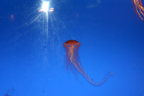
I was unable to take a picture of the setae so instead, I found a picture that the setae is clearly visble.

7. The earthworm's crops store the worm's food as it comes down the esophagus and the gizzard grinds it down. This process makes the worm well suited to its environment.
Here is a picture that shows the esophagus and gizzard.

8. If I cut past segment 32, I would see the rest of the digestive system, nephridia and the worm's food.
This picture shows a portion of the bottom half of the worm. It shows the digestive system and some nephridia.

9. When earthworms start to reproduce, they both exchange sperm with one another because they are hermaphroditic. Both worms would then proceed to collect the sperm in special sacs where it will stay there until it is mature. Once this happens, the worm's clitellium begins to create a sticky substance that will form a ring which contains the now fertilized eggs.
This picture shows the worm's seminal vesicles.

Tuesday, November 27, 2012

Platyhelminthes


Genus : Maiazoon
Species : Cf. Orsaki
Family : Pseudocecotidae
Order : Polycladida
Date Identified : May 25, 1012


Genus: Pseudoceros
Species : Sp.
Family : Pseudocerotidae
Order: Polyclodida
Date Identified : April/May 2002
Length : About 10cm


Genus: Pseudobiceros
Species: Gratus
Family: Pseudocerotidae
Order: Polycladida
Date Identified: 4/2001
Length: 25mm

Sunday, November 25, 2012

Aquarium Field trip

            On Thursday, the class all went to the much anticipated aquarium field trip! Prior to the field trip, everyone was talking about how exciting the aquarium was going to be. Upon arriving, we were separated according to our block to divide us into our stations. Our block was sent to do the marine lab first. During this lab, I learned a lot about different types of sea critters and their habitats. When examining the different types of organisms, our instructor let us touch the various organisms. From sea urchins to anemones, we learned about their role in the environment and why they were so important. Being able to physically pick up the organisms was my favorite part of this field trip because personally, I am fascinated by marine life. After the short hour and a half, we moved on towards doing some experiments with sea snails. This part of the lab was not as exciting as the other, but it was still interesting to learn about the sea snails and performing experiments. In our group, we tested if the snail could swim; which was proven false quickly, and we also tested if the snails were able to flip themselves if their shell was placed on the ground. In our group, the only time they were able to flip over was when they had a rock nearby so they could use that as leverage. After our marine lab was done, we all left to go and have lunch, which was expensive. Shortly after we finished eating, everyone split up and went into the aquarium to search for the answers to the many questions that we had to answer. Some questions were a lot harder to answer than others; however, with the help of some friends, I managed to successfully answer all of the questions. When exploring the aquarium, it was really interesting to take a look at the diversity of all of the organisms inhabiting our oceans. Reading the little information boards were very educational in showing us the role of that particular organism and a lot of them displayed an interesting fact. My favorite organism that I found in the aquarium was the jellyfish. I have always been fascinated with them because they are so unique to everything else which was perfect because we had just learned about them a day or two before hand. This field trip was one of my personal favorites because not only did we get to look at the different types of sea creatures through the glass, we were able to physically touch some of them. This field trip was very educational in the sense that we got to learn the way we wanted to. We got to hands on learn about organisms, or we could read the information displayed all around the aquarium. I only wish all field trips could be this exciting.

1.  The symbiotic relationship between the Clown Anemone Fish and the sea anemones is fairly simple. Since the clown fish have very similar cells to the anemone, they cannot be stung by the anemone's tentacles. In doing so, the clown fish can use the sea anemone as a place for shelter. In return, animals who prey on the clown fish will be drawn towards the anemone and will be stopped in it's path by the anemone's stinging tentacles. In short, the clown fish receives a home and the anemone receives cleaning from the clown fish and food,

2. It is so critical for a sea otter to have a clean pelt of fur because only clean fur is capable or trapping air. Since the otters have no blubber layer to keep them warm, they depend on this air coat to be a barrier between the water and the skin.

3.  Before the 1900s,  sea otters could be found near Amchtka Island and Alaska. They were hunted to near extinction during the fur trades, so some otters were taken from Alaska in order to breed them and repopulate the species in 1969- 1970.

4. The sea anemones that were located beside the sea otter tank was a light green.


5. My favorite invertebrate that was in the aquarium had to be the Japanese Sea Nettle. It got it's name because they throw a powdered sea nettle on an attacker's face which makes them 'sneeze'.


6.  The scientific name for a beluga whale is Delphinaplerus Leucus. In the Vancouver aquarium, there are two currently living there.

7.  According to the aquarium staff, the potbelly sea horse was removed from the aquarium, so we found another species of seahorse. It moves by slowly using it's tail to propel itself forward.

8.  The organism that closely represents the human nervous system is the Pacific Sea Nettle because it's tentacles could represent the nerves in the body  and the head of the jellyfish would represent our head and all of our nervous system sprouts from.

9. The Starry flounder is unique to other fish because of it's flat appearance. Since it is flat, it is able to lurk in the sand to hide from predators or catch their prey and because of it's colour, it camouflages well with the ocean floor. 

10. The Moon-Jellies move slowly by propelling their 'head' by contracting and relaxing. A way to describe their movement would be that they waft through the water. The Moon-Jelliies  contract and expand their head and they push water and plankton into their mouth which is how they eat.


11. The Pacific Octopus has white sunction cups on all of it's arms because they can be used to grapple onto prey or surfaces. This allows them to climb through small crevices. It is considered one of nature's best predators because of it's ability to hide.

12.  The Stellar Sea Lions eat fish and squid and hunt like most mammals do. To hunt prey, they chase down an organism in large groups.

13. The Caiman can be found in Brazil, Argentina, Bolivia and Paraguay. 

14.  The colour of the crocodile is a brown grayish colour.  This colour is perfect for the environment that it inhabits because it can camouflage with it's environment.

15.  The largest fish in the aquarium is the Arapaima. It can grow up to almost three meters long.

16. The vegetarian fish that inhabited the Amazon flooded forest is the Redhook Silver Dollar. It eats the seeds off of riverside shrubs. 

17.  Piranhas are the biggest threat to humans when we have to removing a hook in the mouth.

18. Sharks do not have any bones in their body, instead they have cartilage

19. Some of the other organisms that were living in the tank with the shark were Green Sea Turle, Blacktip Reef Shark, White Spotted Bamboo shark, Stringray, Laced Moray, Common Bluestripe Snapper, Redtail Butterfly fish, Yellowface Angelfish and the Indo-Pacific Sergeant.
20.  The Giant Red Sea Urchin's scientific name is the Strongylocentrotus Francisconus. The urchin has a body surrounded by spikes to protect it's body.

21. The Archerfish shoots a jet of water out at insects and when the insect falls into the water, they eat it.

22. The scientific name of the sea turtle in the tropic zone is the Chelonia Mydas, but people at the aquarium gave her the name 'Schoona'.